
Contents
| Image | Title | Category | Type | Description | Updated |
|---|---|---|---|---|---|
|
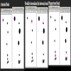
Semi-automated quantification of three stages of phagocytosi using ImageJ | Software | Workflow | The authors present an ImageJ-based, semi-automated phagocytosis workflow to rapidly quantitate three distinct stages during the early engulfment of opsonized beads. |
05/17/2023 - 16:27 |
|
FluoGAN | Software | Component | FluoGAN is a fluorescence image deconvolution software combining the knowledge of acquisition physical model with gan. It takes a fluctuating sequence of blurred, undersampled and noisy images of the sample of interest fixed sample as input from wide field or confocal and returns a super resolved image. |
03/31/2023 - 10:44 |
|
ShareLoc | Dataset | Dataset of single molecule localisation microscopy SMLM, mainly storm, d-storm, dna-paint for now. Data are mainly the localisation positions in text files, some are associated with brightfield images. https://doi.org/10.1038/s41592-022-01659-0
|
05/03/2023 - 11:11 | |
|
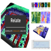
Relate | Software | Collection |
Relate is a correlative software package optimised to work with EM, EDS, EBSD, & AFM data and images. It provides the tools you need to correlate data from different microscopes, visualise multi-layered data in 2D and 3D, and conduct correlative analyses.
|
05/17/2023 - 16:38 |
|
LIVECell | Dataset | LIVECell is a manually annotated and expert-validated dataset of 2D phase contrast images, consisting of over 1.6 million cells from a diverse set of cell morphologies and culture densities. It is also associated with some trained models. All are published under CC BY-NC 4.0 license. |
03/28/2023 - 12:47 | |
|
Junction Mapper | Software | Workflow | Junction Mapper is a semi-automated software (Java Desktop application) for analysing data from images of cells in close proximity to each other in monolayers. The focus of Junction Mapper is to measure the morphology of cell boundaries, define single junctions and quantify the length, area and intensity of the staining of different proteins localised at cell-cell contacts. The output are various unique parameters that assess the contacting interface between cells and up to two junctional markers. |
04/27/2023 - 12:36 |
|
SynActJ | Software | Workflow | SynActJ (Synaptic Activity in ImageJ) is an easy-to-use fully open-source workflow that enables automated image and data analysis of synaptic activity. The workflow consists of a Fiji plugin performing the automated image analysis of active synapses in time-lapse movies via an interactive seeded watershed segmentation that can be easily adjusted and applied to a dataset in batch mode. The extracted intensity traces of each synaptic bouton are automatically processed, analyzed, and plotted using an R Shiny workflow. |
03/22/2023 - 09:39 |
|
Point Cloud Analyst (PoCA) | Software | Collection | SMLM is a mature but still growing field, which still lacks efficient and user-friendly analysis and visualization software platform adapted for both users and developers. We here introduce PoCA, a powerful open-source software platform dedicated to the visualization and analysis of 2D and 3D point-cloud data. |
05/11/2023 - 13:03 |
|
Nanotomy: Large-scale electron microscopy (EM) datasets | Dataset | Nanotomy shares based on the ATLASTM browser-based viewer from Zeiss. This database allow to browse data set from the publications of Giepmans lab. The full list of data set is availble from : Except the papers and where otherwise noted, this work is licensed under a Creative Commons Attribution 4.0 International License
Images can be downloaded but only as screenshots (saved as png). |
03/14/2023 - 12:46 | |
|
Orthanc | Software | Component | Orthanc aims at providing a simple, yet powerful standalone DICOM server. It is designed to improve the DICOM flows in hospitals and to support research about the automated analysis of medical images. Orthanc lets its users focus on the content of the DICOM files, hiding the complexity of the DICOM format and of the DICOM protocol. |
03/14/2023 - 12:32 |
|
SNEMI3D: 3D Segmentation of neurites in EM images | Dataset | In this challenge, a full stack of electron microscopy (EM) slices will be used to train machine-learning algorithms for the purpose of automatic segmentation of neurites in 3D. This imaging technique visualizes the resulting volumes in a highly anisotropic way, i.e., the x- and y-directions have a high resolution, whereas the z-direction has a low resolution, primarily dependent on the precision of serial cutting. |
03/06/2023 - 16:50 | |

|
Cell-IDR | Dataset | The Image Data Resource (IDR) is a public repository of image datasets from published scientific studies, where the community can submit, search and access high-quality bio-image data. It is part of The BioImage Archive stores and distributes biological images that are useful to life-science researchers. Its development will provide data archiving services to the broader bioimaging database community. This includes added-value bioimaging data resources such as EMPIAR, Cell-IDR and Tissue-IDR. |
03/06/2023 - 16:44 | |

|
Tissue-IDR | Dataset | The Image Data Resource (IDR) is a public repository of image datasets from published scientific studies, where the community can submit, search and access high-quality bio-image data. It is part of The BioImage Archive stores and distributes biological images that are useful to life-science researchers. Its development will provide data archiving services to the broader bioimaging database community. This includes added-value bioimaging data resources such as EMPIAR, Cell-IDR and Tissue-IDR.
|
03/06/2023 - 16:40 | |

|
EMPIAR | Dataset | EMPIAR (https://empiar.org/ or https://www.ebi.ac.uk/empiar), the Electron Microscopy Public Image Archive, is a public resource for raw images underpinning 3D cryo-EM maps and tomograms (themselves archived in EMDB). EMPIAR also accommodates 3D datasets obtained with volume EM techniques and soft and hard X-ray tomography. |
03/06/2023 - 16:29 | |

|
Cytomine Open Collection | Dataset | The Cytomine cooperative host several virtual slides having an interest for pedagogical purposes or technical interest. |
03/06/2023 - 16:23 | |
| |
CXIDB | Dataset | The main goal of the Coherent X-ray Imaging Data Bank is to create an open repository for CXI experimental data. |
03/06/2023 - 16:15 | |
| |
Cell Image Library | Dataset | The Cell Image library is a large repository of images from all organisms, cell types, and processes, normal and pathological. Original format and ome-tiff format can be downloaded. |
03/06/2023 - 16:09 | |
|
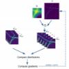
Correlia | Software | Component | Correlia is an open-source ImageJ/FIJI plug-in for the registration of 2D multi-modal microscopy data-sets. The software is developed at ProVIS - Centre for Correlative Microscopy and is specifically designed for the needs of chemical microscopy involving various micrographs as well as chemical maps at different resolutions and field-of-views. |
03/06/2023 - 15:17 |
|
empanada-napari | Software | Component | The empanada-napari plugin is built to democratize deep learning image segmentation for researchers in electron microscopy (EM). It ships with MitoNet, a generalist model for the instance segmentation of mitochondria. There are also tools to quickly build and annotate training datasets, train generic panoptic segmentation models, finetune existing models, and scalably run inference on 2D or 3D data. To make segmentation model training faster and more robust, CEM pre-trained weights are used by default. |
01/25/2023 - 15:51 |

|
MIA | Software | Collection | ModularImageAnalysis (MIA) is an ImageJ plugin which provides a modular framework for assembling image and object analysis workflows. Detected objects can be transformed, filtered, measured and related. Analysis workflows are batch-enabled by default, allowing easy processing of high-content datasets. |
08/14/2023 - 12:43 |