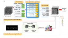
Free and open source

Description

The Allen Cell Structure Segmenter is a Python-based open source toolkit developed at the Allen Institute for Cell Science for 3D segmentation of intracellular structures in fluorescence microscope images.

It consists of two complementary elements:

  1. Classic image segmentation workflows for 20 distinct intracellular structure localization patterns. A visual “lookup table” is outlining the modular algorithmic steps for each segmentation workflow. This provides an intuitive guide for selection or construction of new segmentation workflows for a user’s particular segmentation task. 
  2. Human-in-the-loop iterative deep learning segmentation workflow trained on ground truth manually curated data from the images segmented with the segmentation workflow. Importantly, this module was not released yet.


The Allen Cell Structure Segmenter Overview
Description

DeconvolutionLab2 includes a friendly user interface to run the following deconvolution algortihms: Regularized Inverse Filter, Tikhonov Inverse Filter, Naive Inverse Filter, Richardson-Lucy, Richardson-Lucy Total Variation, Landweber (Linear Least Squares), Non-negative Least Squares, Bounded-Variable Least Squares, Van Cittert, Tikhonov-Miller, Iterative Constraint Tikhonov-Miller, FISTA, ISTA.

The backbone of our software architecture is a library that contains the number-crunching elements of the deconvolution task. It includes the tool for a complete validation pipeline. Inquisitive minds inclined to peruse the code will find it fosters the understanding of deconvolution.

has topic
has function
Description

quote: 

GaussFit_OnSpot is an ImageJ plugin for fitting Gaussian profiles onto selected positions in diffraction-limited images (e.g. single molecules, protein clusters, vesicles, or stars).

The plugin performs a function fit in regions of interest (ROI) around spots marked by point selections in grayscale images. Single or multiple spots can be either selected manually with the Multi-point tool or automatically with the Find Maxima function.

There is a PDF with more information, and also an example image.

has function
Description

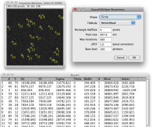
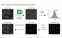
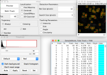
Quote " finding and/or analyzing colocalization of bright intensity spots (cells, particles, vesicles, comets, dots, etc) in images with heterogeneous background (microscopy, astronomy, engineering, etc). "

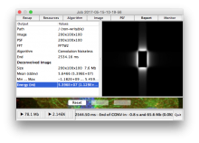
Uses Gaussian-Mexican hat convolution for preprocessing.

Description

"PTA2 is an ImageJ1.x plugins that enable automatic particle tracking"

This plugin is developed specifically for single-molecule imaging, so it's good at tracking spots with noisy background. 

has function