Python

Description

Orthanc aims at providing a simple, yet powerful standalone DICOM server. It is designed to improve the DICOM flows in hospitals and to support research about the automated analysis of medical images. Orthanc lets its users focus on the content of the DICOM files, hiding the complexity of the DICOM format and of the DICOM protocol.

Orthanc can turn any computer running Windows, Linux or OS X into a DICOM store (in other words, a mini-PACS system). Its architecture is lightweight and standalone, meaning that no complex database administration is required, nor the installation of third-party dependencies.

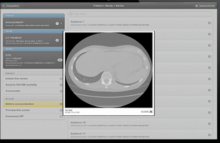
What makes Orthanc unique is the fact that it provides a RESTful API. Thanks to this major feature, it is possible to drive Orthanc from any computer language. The DICOM tags of the stored medical images can be downloaded in the JSON file format. Furthermore, standard PNG images can be generated on-the-fly from the DICOM instances by Orthanc.

Orthanc also features a plugin mechanism to add new modules that extends the core capabilities of its REST API. A Web viewer, a PostgreSQL database back-end, a MySQL database back-end, and a reference implementation of DICOMweb are currently freely available as plugins.

orthanc
Description

The empanada-napari plugin is built to democratize deep learning image segmentation for researchers in electron microscopy (EM). It ships with MitoNet, a generalist model for the instance segmentation of mitochondria. There are also tools to quickly build and annotate training datasets, train generic panoptic segmentation models, finetune existing models, and scalably run inference on 2D or 3D data. To make segmentation model training faster and more robust, CEM pre-trained weights are used by default. These weights were trained using an unsupervised learning algorithm on over 1.5 million EM images from hundreds of unique EM datasets making them remarkably general.

Empanada-napari
Description

ASTEC stands for Adaptive Segmentation and Tracking of Embryonic Cells. It proposes a full workflow for time lapse light sheet imaging analysis, including drift/motion compensation before the segmentation itself, and the capacity to correct for it.  It was used to process 3D+t movies acquired by the MuViSPIM light-sheet microscope in particular.

Astec embryon
Description

ClearMap is a toolbox for the analysis and registration of volumetric data from cleared tissues.

It was initially developed to map brain activity at cellular resolution in whole mouse brains using immediate early gene expression. It has since then been extended as a tool for the qunatification of whole mouse brain vascualtur networks at capilary resolution.

It is composed of sevral specialized modules or scripts: tubemap, cellmap, WobblyStitcher.

ClearMap has been designed to analyze O(TB) 3d datasets obtained via light sheet microscopy from iDISCO+ cleared tissue samples immunolabeled for proteins. The ClearMap tools may also be useful for data obtained with other types of microscopes, types of markers, clearing techniques, as well as other species, organs, or samples.

ClearMap SCreenshot
Description

Quote:

pyTFM is a python package that allows you to analyze force generation and stresses in cells, cell colonies, and confluent cell layers growing on a 2-dimensional surface. This package implements the procedures of Traction Force Microscopy and Monolayer Stress Microscopy. In addition to the standard measures for stress and force generation, it also includes the line tension, a measure for the force transfer exclusively across cell-cell boundaries. pyTFM includes an addon for the image annotation tool clickpoints allowing you to quickly analyze and vizualize large datasets.

https://pytfm.readthedocs.io/en/latest/_images/mask_force_measures.png