Description
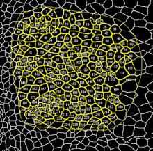
This macro is meant to segment the cells of a multicellular tissue. It is written for images showing highly contrasted and uniformly stained cell membranes. The geometry of the cells and their organization is automatically extracted and exported to an ImageJ results table. This includes: Cell area, major, minor fitted ellipse radii + major axis orientation and number of neighbors of the cells. Manual correction of the automatic segmentation is supported (merge split cells, split merged cells).
Sample image data is available in the documentation page.