
- 254 views
Raw data of three types of dendritic spines imaged in biphoton (mushroom, tubby, thin spines).
Hippocampal neurons from mouse organotypic slice cultures
If you use this dataset , please cite
M. U. Ghani, F. Mesadi, S. D. Kanik, A. O. Argunsah, A. Hobbiss, I. Israely, D. Unay, T. Tasdizen, and M. Cetin, "Shape and appearance features based dendritic spine classification," Journal of Neuroscience Methods.
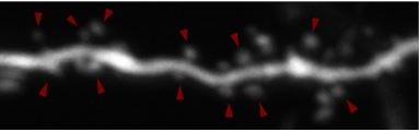
Annotated data and mask labels provided
Two-photon imaging was performed using a galvanometer-based scanning system (Prairie Technologies, acquired by Bruker Inc.) on an Olympus BX61WI equipped with 60X water immersion objective (0.9 NA), using a Ti:sapphire laser (Coherent Inc.) controlled by PrairieView software at 910 nm. Z-stacks (0.3 μm axial spacing) from secondary or tertiary dendrites from CA1 neurons were collected every 5 min for up to 4 h. The field of view was 19.8 × 19.8 μm at 1024 × 1024 pixels.