Light microscopy

Synonyms
Optical microscopy
Optical microscope
Light microscope
Description

ClearVolume is a real-time live 3D visualization library designed for high-end volumetric microscopes such as SPIM and DLSM microscopes. With ClearVolume you can see live on your screen the stacks acquired by your microscope instead of waiting for offline post-processing to give you an intuitive and comprehensive view on your data. The biologists can immediately decide whether a sample is worth imaging. ClearVolume can easily be integrated into existing Java, C/C++, Python, or LabVIEW based microscope software. It has a dedicated interface to MicroManager/OpenSpim/OpenSpin control software. ClearVolume supports multi-channels, live 3D data streaming from remote microscopes, and uses a multi-pass Fibonacci rendering algorithm that can handle large volumes. Moreover, ClearVolume is integrated into the Fiji/ImageJ2/KNIME ecosystem. You can now open your stacks with ClearVolume from within these popular frameworks for offline viewing.

has function
Description

Microscopy Image Browser (MIB) is a high-performance Matlab-based software package for advanced image processing, segmentation and visualization of multi-dimensional (2D-4D) light and electron microscopy datasets.

MIB is a freely available, user-friendly software for effective image processing of multidimensional datasets that improves and facilitates the full utilization of acquired data and enables quantitative analysis of morphological features. Its open-source environment enables fine tuning and possibility of adding new plug-ins to customize the program for specific needs of any research project.

MIB
Description

iTrack4U is a Java-based software using ImageJ and jMathPlot libraries, which aims at automatically tracking cells recorded in phase-contrast microscopy. It includes all tools from image files preprocessing, tracking to data extraction and visualization. 


Please cite Cordeliéres et. al. (2013) when using this software package!

iTrack4U
Description

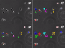
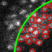
CIDRE is a retrospective illumination correction method for optical microscopy. It is designed to correct collections of images by building a model of the illumination distortion directly from the image data. Larger image collections provide more robust corrections. Details of the method are described in

K. Smith, Y. Li, F. Ficcinini, G. Csucs, A. Bevilacqua, and P. Horvath
CIDRE: An Illumination Correction Method for Optical Microscopy, Nature Methods 12(5), 2015, doi:10.1038/NMETH.3323

Illumination correction method
Description

Spot detector detects and counts spots, based on wavelet transform.

- Detects spots in noisy images 2D/3D.
- Depending on objective, spots can be nuclei, nucleus or cell
- Versatile input: sequence or batch of file.
- Detects spot in specific band/channel.
- Multi band labeling: automaticaly creates ROIs from one band and count in the same or an other band.
- Filters detection by size.
- Sort detection by ROIs
- Output data in XLS Excel files: number of detection by ROIs, and each detection location and size.
- Outputs withness image with ROIs and detection painted on it.
- Outputs binary detection image.
- Displays detections
- Displays tags

logo spot detector